+919849193447

know more about

orbital Implants

A know how

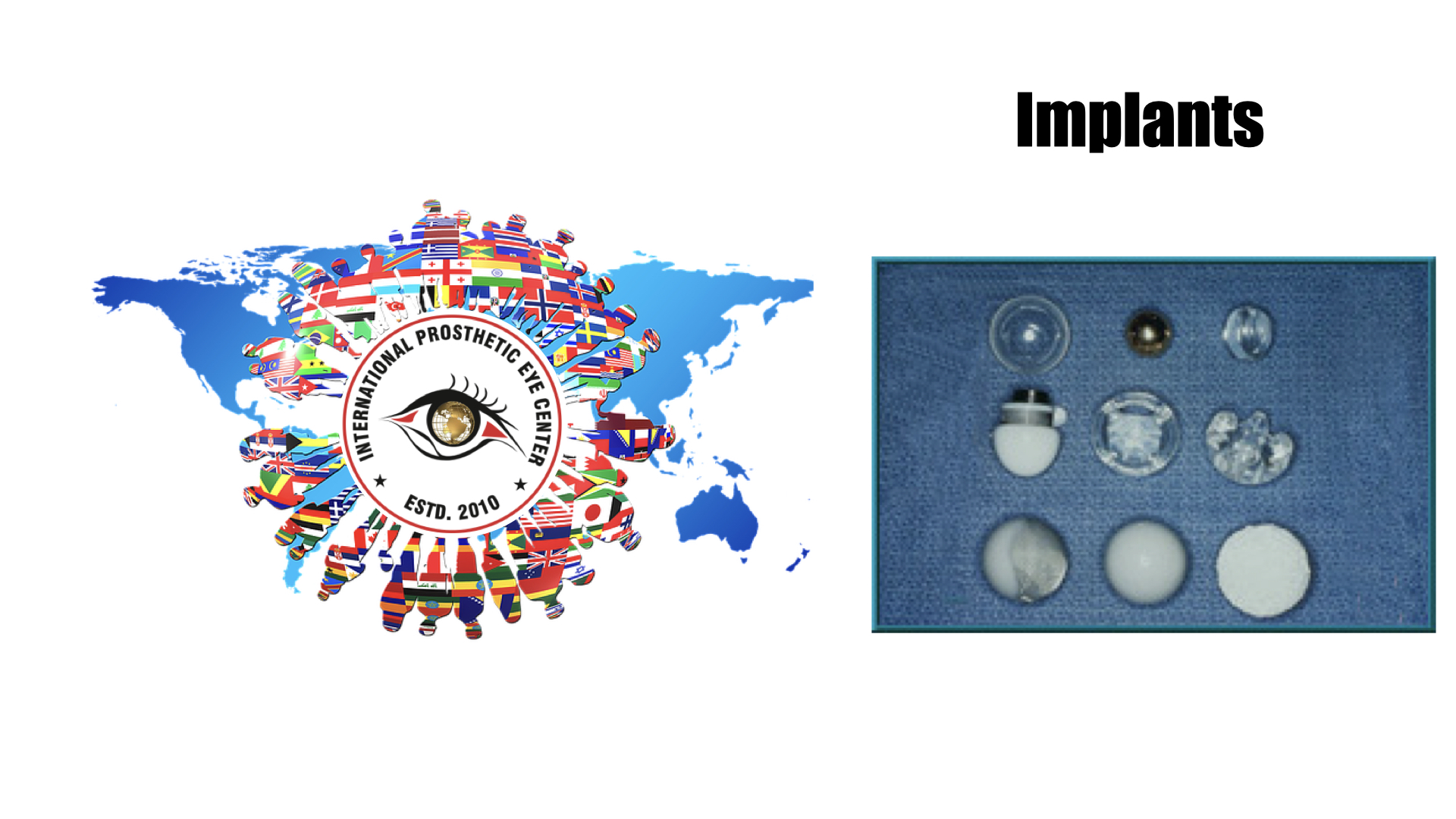
There are several types of implants that may be utilized in an enucleation. Implants are made in distinct sizes, and intraoperative selection of the appropriately sized implant is determined by the size of the patient’s orbit and the size of the implant necessary to achieve symmetry with the fellow eye. Implants may be porous or nonporous. Porous implants allow for anchoring of the extraocular muscles with proliferation of fibrovascular tissues into the implant itself. These include hydroxyapatite, porous polyethylene, and proplast. Some considerations regarding time of implant can include presence of infection and/or fracture(s).

Hydroxyapatite implants were first introduced in 1989.[9] Because of their rough surface, they are typically wrapped with material such as donor sclera, acellular dermis, or pericardium. Additional wrapping materials include autologous tissue grafts, such as temporalis fascia or fascia lata and synthetic meshes. The extraocular muscles may be then sutured to the wrapping material for enhanced motility of the implant. Porous polyethylene implants were later developed as an alternative to hydroxyapatite. They have a smoother surface and do not require wrapping. Other advantages of porous polyethylene over hydroxyapatite implants include cheaper cost and ability to suture the extraocular muscles directly to the implant.[3]

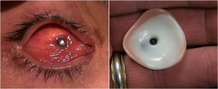
Figure 6: Pegged implant and prosthesis.

Implants may be pegged, in which a hole is drilled into the implant where a peg can be placed that attaches to the prosthesis. Pegging is typically performed six to twelve months postoperatively and allows for increased motility of the prosthesis.[3] Compared to unpegged porous implants, pegged orbital implants were found in one study to produce a statistically significant improvement in horizontal but not vertical motility.[10] Pegged implants were found to retain 87% of motility of the fellow eye while unpegged implants were found to retain only 50% of motility of the fellow eye.

In one series of 802 patients who underwent evisceration, enucleation, or placement of a secondary implant where a hydroxyapatite implant was used, 156 of 353 (44%) of patients with pegged implants had a complication. The most common complication in this series was peg extrusion in 20% of cases, which was shown to occur less frequently with titanium pegs as opposed to other types of pegs.[11] In another retrospective series of patients with pegged hydroxyapatite implants, complications were noted in 38% (62/165) of patients. Complications of pegging included discharge (most common), pyogenic granulomas, loss of peg, poor transfer of movement, and audible click.[12] Two patients (3%) had implant infection that required removal of the implant. Because of these complications, pegging has fallen out of favor and is now utilized less commonly.

Nonporous implants do not allow for proliferation of tissues into the implant. Therefore, they may have decreased motility and greater risk of implant migration, yet several studies indicate no problems with implant migration with appropriate technique. [13][14]Types of nonporous implants include glass, silicone, acrylic, and polymethylmethacrylate (PMMA).[3] The rectus muscles still may be sutured over the implant to impart motility to the implant and prosthesis, or suture to a wrapped material.

In a survey published in 2004 of active members of the American Society of Ophthalmic Plastic and Reconstructive Surgery (ASOPRS) regarding management trends and preferences in primary enucleations and eviscerations, the most popular and commonly used implant was porous polyethylene in 43% of cases, followed by hydroxyapatite (27%) and nonporous implants (20%).[15] The most commonly cited rationale for implant choice was outcome. Pegs were utilized in only 8% of cases. A majority of implants were not wrapped, but, of those that were, donor sclera was the most commonly utilized wrapping material.

Postoperative Management

Patients are typically patched for a brief period postoperatively and instructed to return to clinic one week after the procedure. Prescriptions are given for analgesics and antiemetics. Some surgeons also elect to prescribe prophylactic antibiotics, although there is no evidence that antibiotics reduce the risk of implant infection.[16][17][18]   

Once the conjunctiva closure has healed, generally about 4-8 weeks postoperatively, patients are referred to an ocularist for fitting of an ocular prosthetic fitting. Patients will require regular follow up with both an oculoplastic surgeon and an ocularist to maintain the health of their socket.


Complications

Complications of an enucleation include the following:

Intraoperative:

Figure 7: Exposure of PMMA, porous polyethylene, and hydroxyapatite implants (from left to right).

  • Removal of the wrong eye

  • Damage to or loss of extraocular muscles

  • Hemorrhage

  • Perforation of eye


Postoperative:

  • Infection

  • Hemorrhage

  • Wound dehiscence

  • Extrusion of the conformer

  • Contraction of the fornices

  • Exposure of the implant

  • Extrusion of the implant

  • Migration of the implant

  • Ptosis

  • Pain

  • Ectropion

  • Entropion

  • Hollow or deep superior sulcus

  • Poorly fitting prosthesis

  • Enophthalmos

  • Socket contracture

  • Orbital cellulitis

Acknowledgements

Thank you to Oculoplastics Associates of Texas, Dallas, TX for providing article images.

Welcome International Prosthetic Eye Center

Your life-like Customized Artificial Eyes

We offered very natural looking artificial eyes!

In certain cases where normal eye is bulging forward, or have pain in the eyes with out Vision, these patient may under go for evisceration or enucleation with or with out use of orbital implant. The surgical procedure to remove the entire natural eye is referred to as an enucleation where as the surgical procedure to remove the contents of of a natural eye is referred to as evisceration.Both procedures will need prosthetic restoration with the fitting of an artificial eye. This process can usually begin approximately six weeks following the initial surgery.

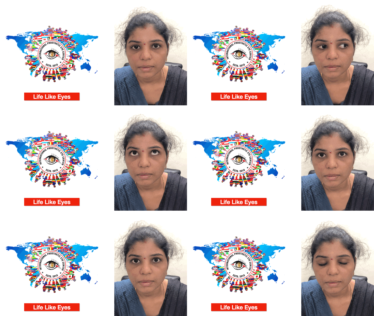
Fitting the artificial eye begins with a board certified ocularist taking an impression mold of the surgically repaired eye socket or residual eye. This procedure ensures each patient attains the maximum amount of comfort and movement possible.

Our team of board certified ocularists have developed advanced art and sculpting techniques to recreate artificial eyes with life-like and natural appearance. These techniques are focus on carefully matching eyelid contours, eyelid folds, and other features involved in achieving facial symmetry. We make every effort to maximize the amount of movement and to recreate an exact color match to your natural eye.

At International Prosthetic Eye Center, we utilize a combination of digital photography and hand painted technques to replicate the color and details of your eye. Only natural earth pigments are used in the painting of our artificial eyes. These pigments provide exquisite detail, depth of color, and vibrancy for the life of your artificial eye.

TOP EXPERT IN INDIA

EXPERT IN providing TREATMENT post Orbital Surgery

Team is highly qualified and very passionated to care for you at every step of providing care.

Kuldeep Raizada, Ph D BCO, BADO, FAAO

Kuldeep Raizada is a Licensed Ocularist in Hyderabad, a Board Certified Ocularist (BCO) from National examination Board of ocularist’s, USA and a Board Approved Diplomate Ocularist (BADO) from American Society of ocularist’s, specializing in ocular prosthetics since 2001, Kuldeep places his emphasis on the satisfaction and well-being of every patient. His clinical skill and expertise are equally matched by his personalized care for patients and attention to detail.

Kuldeep Raizada completed his basic optometry education at Gandhi Eye Hospital, Aligarh, and has his training at L V Prasad Eye Institute, Hyderabad. where he was also Founder and Head of the Department of Ocular Prosthesis services till 2009. He completed a second fellowship, in Anaplastolgy, at MD Anderson Cancer Centre, Houston. He has also been trained by the top most ocularist and anaplastologist in United States of America.

His clinical interests include ocular and facial prosthesis, particularly in pediatric patients. His research interests lie in newer advancement in development of new types of prosthesis, newer solution for ptosis corrective glasses.

Kuldeep Raizada, is Founder & Director of the International Prosthetic Eye Center since 2010, where he is practicing since 2010.

Kuldeep Raizada has been recognized by the American Society of Ocularist, USA and American Anaplastology Association,USA and by several other professional organizations, for his excellence in research and clinical practice.

Kuldeep Raizada, have completed all requirements by American Society of Ocularist, which is hard work of 14000 working hours as well extensive study for prosthetics, Hence awarded the Diplomate Ocularist from American Society of Ocularist, USA, 2012, Chicago, USA, which is the First ever received all over Asia Pacific & throughout Middle East so ever.

At present he is reviewer of several journals like Contact Lens & Anterior Eye, International Journal of Anaplastology, Oculoplasty & Reconstructive Surgery (OPRS) and Many others. He has published and presented world widely.

Fact About Kuldeep

  • CEO & Chairman of Akriti

  • Founder & Director International Prosthetic Eye Center, India

  • Founder of Healthcare India Magazine

  • Receipt of Abdul Kalam Award.

  • First ever Indian to received Board Certifications, from National Board of examination of ocularist, USA, He was the first One all over Asia Pacific & throughout Middle East.

  • Developed First Digital Dynamic Facial Eye Prosthesis

  • Developed physiological Near Stereo test

  • Developed physiological Distance Stereo test

  • Developed ETDRS Log Mar Vision Charts in Hindi for 4 Meters

  • Developed ETDRS Log Mar Vision Charts in Telegu for 4 Meters 

  • Developed ETDRS Log Mar Vision Charts in Assamese for 4 Meters 

  • Developed ETDRS Log Mar Vision Charts in Arabic for 4 Meters 

  • Developed ETDRS Log Mar Vision Charts in Bangla for 4 Meters 

  • Developed ETDRS Log Mar Vision Charts in Tamil for 4 Meters 

  • Developed ETDRS Log Mar Vision Charts in Oriya for 4 Meters 

  • Developed ETDRS Log Mar Vision Charts in Kannada for 4 Meters

  • Developed ETDRS Log Mar Vision Charts in Malayalam for 4 Meters 

Deepa D Raizada, MS, BCA, BCO, BCCA

Deepa completed her diploma and clinical fellowship in optometry at L V Prasad Eye Institute, India, in 2003, pursued her graduation from Madhurai Kamraj University, 2006.

She had completed her Master of Science (M Sc) in “Maxillofacial and Craniofacial Technology” (2010-2012), King’s Collage London, UK where she was trained to work exclusively in the field of Maxillofacial Prosthetics.

Deepa is also an associate Member of The Institute of Maxillofacial Prosthetist & Technologist, UK (AIMPT) since 2014. & Active Member of International Anaplastology Association, (IAA) USA since 2009.

Deepa Have make a remarkable success in becoming "Asia's First Board Certified Clinical Anaplastologist ( BCCA") and Bring glory for India as to Make India Fifth Nation to have a Board Certified Clinical Anaplastologist.

She Did complete her Board Certification for Ocularist (BCO), By National Examination Board of ocularist, USA in March 2018

She persued her basic training in Ocularistry from L V Prasad Eye Institute, Hyderabad and advanced training in Ocular Prosthetics (May – July, 2005) at Moorefield’s Eye Hospital, London, UK under Mr Nigel Saap.

She worked as an Ocularist at Ocular Prosthesis Department, L V Prasad Eye Institute from 2003 – 2010. She worked with different type of techniques, developed new techniques in the fabrication and fitting of ocular & facial Prostheses. Her work was well recognized and appreciated.

Her clinical interest include make her career in the art and science of facial prosthetics and ocular prosthetics, particularly in pediatric patients. Her research interests lie in developing new techniques in the field of facial prosthetics, and undertake research on materials used in this field.

Deepa Raizada has been recognized by the Oculoplasty Society of India, Indian Optometric Association and by several other professional organizations in India as well as Internationally, for her excellence in research and clinical practice.

Email ID: deepadraizada@gmail.com, deepaocularist@gmail.com

Fact about Deepa Raizada:

  1. She is the First Indian Lady to be American Board Certified Ocularist (BCO) as well as Board Certified Clinical Anaplastogist (CCA).

  2. Her credentials are unmatched with any one from Asia, Middle East, Africa & Europe. First Ever Indian Lady to be American Board Certified Ocularist (BCO) 2018

  3. First Ever Indian Lady to received Her Board Certification in Clinical Anaplastology (CCA) 3rd Individual in the World to have CCA and BCO, (Other 2 in USA)

  4. She bring the glory to India for bringing the First Ever CCA & made a land mark for India to 5th country in the world.