+919849193447

Know more

enucleation of the Eye

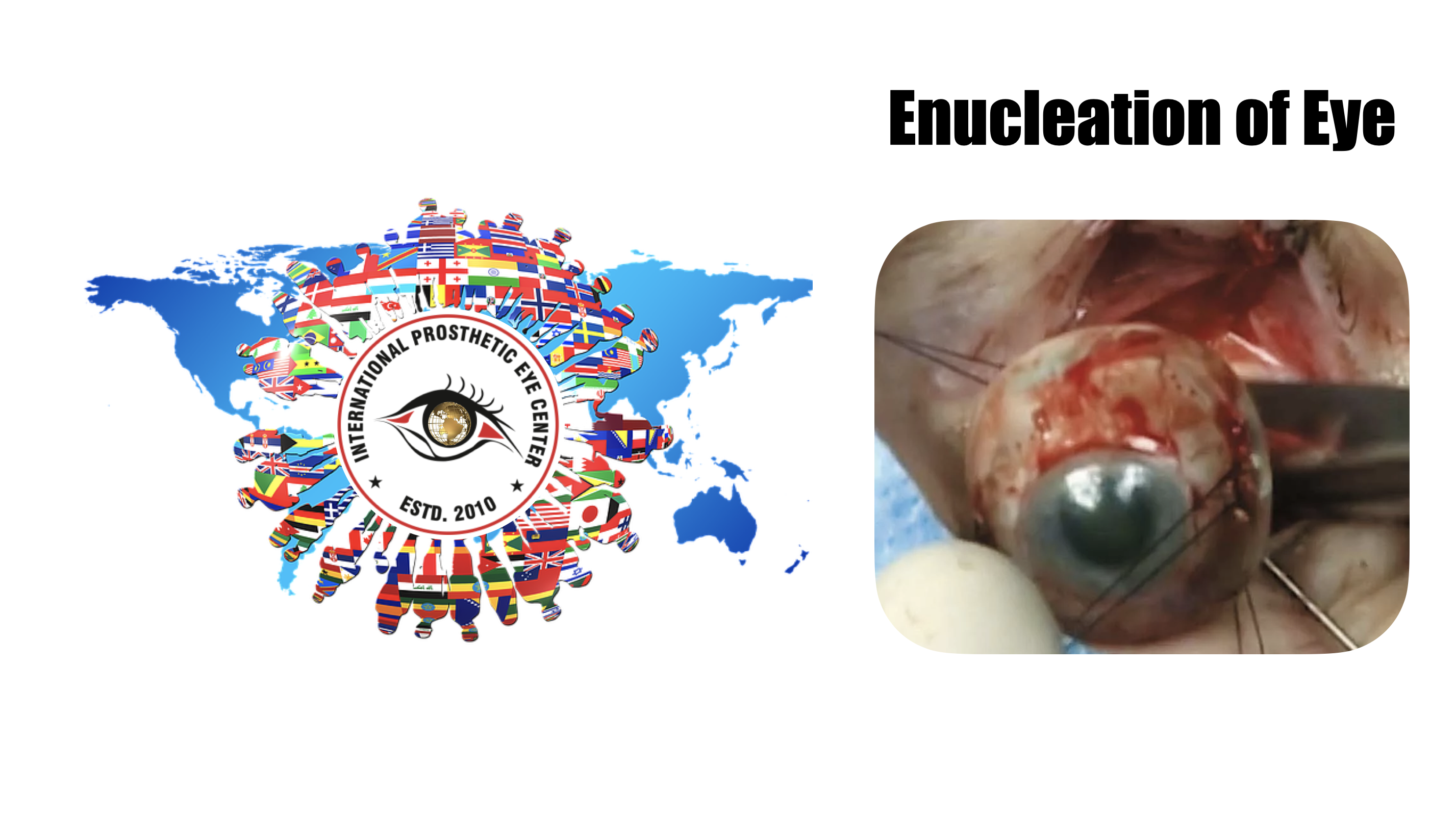
Enucleation is the surgical procedure that involves removal of the entire globe and its intraocular contents, with preservation of all other periorbital and orbital structures. Enucleation is in contrast to evisceration, in which the ocular contents are removed from an intact sclera, and exenteration, in which the entire orbital contents, including the globe and soft tissues, are removed.

Background

The surgical removal of the eye was first reported in the 1500s as a procedure known as extirpation.[1] Unlike an enucleation, the conjunctiva and extraocular muscles were not spared. By the mid 1800s, an enucleation without implant placement was described in the literature. The first reports of implant insertion following enucleation were described in 1886 and 1887, with variable success in implant retention.[1]

Indications

The following are indications for enucleation:

  • Intraocular malignancy or high suspicion for intraocular malignancy (most commonly uveal melanoma and retinoblastoma)

  • Trauma without visual potential

  • Blind, painful eye

  • Severe infection without visual potential

  • Sympathetic ophthalmia

  • Microphthalmos


The role of primary enucleation in acute trauma remains controversial, particularly when a patient's mental status may be altered and/or they are unable to consent. Many surgeons advocate for primary closure of an open globe with later consideration for an enucleation if the eye remains no light perception or becomes painful without vision. Enucleation of an eye can be associated with significant psychological trauma, and the approach of primary globe repair provides the patient to consider their options and the pros and cons of enucleation after the initial trauma and any altered mental status has resolved and provides autonomy to elect this operation in the future. The rare risk of sympathetic ophthalmia in the uninvolved eye must be considered and discussed with the patient. In select cases where the eye is determined to have no visual potential, when repair or at least primary closure of the globe is determined to be impossible, and/or the medical comorbidities of the patient are significant, the surgeon may elect to perform primary enucleation.

Advantages

In contrast to evisceration, enucleation allows for histologic examination of an intact globe and optic nerve. This is particularly important in settings of biopsy-proven or suspected intraocular malignancy, in which it is essential to determine the margins of the malignancy and invasion of the optic nerve, if any.

When comparing the aesthetics of an enucleated socket to an eviscerated socket, one retrospective study showed no statistically significant difference between enucleation and evisceration patients graded by both patients and masked observers.[2]

Enucleation classically has been thought to decrease the risk of sympathetic ophthalmia as it avoids exposure to uveal antigens that may occur during an evisceration.[3] However, more recent studies have reported no cases of sympathetic ophthalmia following evisceration.[4][5]

Disadvantages

A reduction in implant motility is often noted in enucleations. Compared to patients who underwent evisceration, one study found enucleation patients to have statistically significant poorer implant motility.[2] However, no difference in prosthesis motility was noted between evisceration and enucleation patients.


In a 2003 survey of all board-certified ocularists in the United States, 92% reported preferring evisceration to enucleation for a patient who required removal of the eye.[6] Eighty-two percent of survey respondents believed that evisceration afforded the best ocular motility and best overall cosmetic outcome, and 94% percent believed that complications of enophthalmos and/or deep superior sulcus were more common after enucleations.

Surgical Technique

An enucleation is often performed as an outpatient procedure under general anesthesia. A retrobulbar block of local anesthetic with epinephrine is administered to aid in hemostasis and postoperative pain management. After a time-out is performed to confirm the correct operative eye with the entire operating room team, the face is prepared and draped in sterile fashion. A limbal conjunctival peritomy is performed with Wescott scissors for 360 degrees. Blunt dissection in the sub-Tenon's plane is then carried out in each of the oblique quadrants. Each rectus muscle is then identified, isolated with a muscle hook, secured with suture, and cut at the insertion to the globe. The superior and inferior oblique muscles are isolated and generally transected, though some surgeons prefer to tag these as well. Conversely, some surgeons prefer to secure the muscles with suture after the eye has been removed.[7] 


Once the globe is determined to rotate freely, the optic nerve is identified, strummed, and cut with enucleation scissors or an enucleation snare wire. Some surgeons prefer to first clamp the optic nerve with a curved hemostat prior to transection to encourage further hemostasis. An attempt should be made to cut a long segment of the optic nerve, particularly in situations of intraocular malignancy where histologic examination of the optic nerve is crucial. Additional hemostasis is then achieved with direct pressure in the intraconal space and cautery of the optic nerve if needed.

An implant is then placed in the intraconal space to replace volume lost by the enucleated globe, achieve cosmetic symmetry with the fellow socket, and allow for motility of the prosthesis. To determine appropriate diameter of the implant, use of the formula axial length-2 mm has been shown to provide for adequate replacement of lost volume and minimize superior sulcus deformity and enophthalmos.[8] A sizing set can also be used to determine the appropriate size intraoperatively.

In certain circumstances including severe infection, a surgeon may choose not to place an implant at the time of enucleation and elect to place an implant in a second surgery. The extraocular muscles are generally attached to each other anterior to the implant or sutured directly to a porus or wrapped implant. A two-layered closure is then carried out with absorbable sutures, first of Tenon’s capsule and then of the conjunctiva. Antibiotic ointment is applied, a clear plastic conformer is placed over the closed conjunctiva, and a pressure patch is placed over the socket. A temporary tarsorrhaphy may be placed.


Welcome International Prosthetic Eye Center

Your life-like Customized Artificial Eyes

We offered very natural looking artificial eyes!

In certain cases where normal eye is bulging forward, or have pain in the eyes with out Vision, these patient may under go for evisceration or enucleation with or with out use of orbital implant. The surgical procedure to remove the entire natural eye is referred to as an enucleation where as the surgical procedure to remove the contents of of a natural eye is referred to as evisceration.Both procedures will need prosthetic restoration with the fitting of an artificial eye. This process can usually begin approximately six weeks following the initial surgery.

Fitting the artificial eye begins with a board certified ocularist taking an impression mold of the surgically repaired eye socket or residual eye. This procedure ensures each patient attains the maximum amount of comfort and movement possible.

Our team of board certified ocularists have developed advanced art and sculpting techniques to recreate artificial eyes with life-like and natural appearance. These techniques are focus on carefully matching eyelid contours, eyelid folds, and other features involved in achieving facial symmetry. We make every effort to maximize the amount of movement and to recreate an exact color match to your natural eye.

At International Prosthetic Eye Center, we utilize a combination of digital photography and hand painted technques to replicate the color and details of your eye. Only natural earth pigments are used in the painting of our artificial eyes. These pigments provide exquisite detail, depth of color, and vibrancy for the life of your artificial eye.

TOP EXPERT IN INDIA

EXPERT IN TREATMENT for Enucleation of eye

Team is highly qualified and very passionated to care for you at every step of providing care.

Kuldeep Raizada, Ph D BCO, BADO, FAAO

Kuldeep Raizada is a Licensed Ocularist in Hyderabad, a Board Certified Ocularist (BCO) from National examination Board of ocularist’s, USA and a Board Approved Diplomate Ocularist (BADO) from American Society of ocularist’s, specializing in ocular prosthetics since 2001, Kuldeep places his emphasis on the satisfaction and well-being of every patient. His clinical skill and expertise are equally matched by his personalized care for patients and attention to detail.

Kuldeep Raizada completed his basic optometry education at Gandhi Eye Hospital, Aligarh, and has his training at L V Prasad Eye Institute, Hyderabad. where he was also Founder and Head of the Department of Ocular Prosthesis services till 2009. He completed a second fellowship, in Anaplastolgy, at MD Anderson Cancer Centre, Houston. He has also been trained by the top most ocularist and anaplastologist in United States of America.

His clinical interests include ocular and facial prosthesis, particularly in pediatric patients. His research interests lie in newer advancement in development of new types of prosthesis, newer solution for ptosis corrective glasses.

Kuldeep Raizada, is Founder & Director of the International Prosthetic Eye Center since 2010, where he is practicing since 2010.

Kuldeep Raizada has been recognized by the American Society of Ocularist, USA and American Anaplastology Association,USA and by several other professional organizations, for his excellence in research and clinical practice.

Kuldeep Raizada, have completed all requirements by American Society of Ocularist, which is hard work of 14000 working hours as well extensive study for prosthetics, Hence awarded the Diplomate Ocularist from American Society of Ocularist, USA, 2012, Chicago, USA, which is the First ever received all over Asia Pacific & throughout Middle East so ever.

At present he is reviewer of several journals like Contact Lens & Anterior Eye, International Journal of Anaplastology, Oculoplasty & Reconstructive Surgery (OPRS) and Many others. He has published and presented world widely.

Fact About Kuldeep

  • CEO & Chairman of Akriti

  • Founder & Director International Prosthetic Eye Center, India

  • Founder of Healthcare India Magazine

  • Receipt of Abdul Kalam Award.

  • First ever Indian to received Board Certifications, from National Board of examination of ocularist, USA, He was the first One all over Asia Pacific & throughout Middle East.

  • Developed First Digital Dynamic Facial Eye Prosthesis

  • Developed physiological Near Stereo test

  • Developed physiological Distance Stereo test

  • Developed ETDRS Log Mar Vision Charts in Hindi for 4 Meters

  • Developed ETDRS Log Mar Vision Charts in Telegu for 4 Meters 

  • Developed ETDRS Log Mar Vision Charts in Assamese for 4 Meters 

  • Developed ETDRS Log Mar Vision Charts in Arabic for 4 Meters 

  • Developed ETDRS Log Mar Vision Charts in Bangla for 4 Meters 

  • Developed ETDRS Log Mar Vision Charts in Tamil for 4 Meters 

  • Developed ETDRS Log Mar Vision Charts in Oriya for 4 Meters 

  • Developed ETDRS Log Mar Vision Charts in Kannada for 4 Meters

  • Developed ETDRS Log Mar Vision Charts in Malayalam for 4 Meters 


Deepa D Raizada, MS, BCA, BCO, BCCA

Deepa completed her diploma and clinical fellowship in optometry at L V Prasad Eye Institute, India, in 2003, pursued her graduation from Madhurai Kamraj University, 2006.

She had completed her Master of Science (M Sc) in “Maxillofacial and Craniofacial Technology” (2010-2012), King’s Collage London, UK where she was trained to work exclusively in the field of Maxillofacial Prosthetics.

Deepa is also an associate Member of The Institute of Maxillofacial Prosthetist & Technologist, UK (AIMPT) since 2014. & Active Member of International Anaplastology Association, (IAA) USA since 2009.

Deepa Have make a remarkable success in becoming "Asia's First Board Certified Clinical Anaplastologist ( BCCA") and Bring glory for India as to Make India Fifth Nation to have a Board Certified Clinical Anaplastologist.

She Did complete her Board Certification for Ocularist (BCO), By National Examination Board of ocularist, USA in March 2018

She persued her basic training in Ocularistry from L V Prasad Eye Institute, Hyderabad and advanced training in Ocular Prosthetics (May – July, 2005) at Moorefield’s Eye Hospital, London, UK under Mr Nigel Saap.

She worked as an Ocularist at Ocular Prosthesis Department, L V Prasad Eye Institute from 2003 – 2010. She worked with different type of techniques, developed new techniques in the fabrication and fitting of ocular & facial Prostheses. Her work was well recognized and appreciated.

Her clinical interest include make her career in the art and science of facial prosthetics and ocular prosthetics, particularly in pediatric patients. Her research interests lie in developing new techniques in the field of facial prosthetics, and undertake research on materials used in this field.

Deepa Raizada has been recognized by the Oculoplasty Society of India, Indian Optometric Association and by several other professional organizations in India as well as Internationally, for her excellence in research and clinical practice.

Email ID: deepadraizada@gmail.com, deepaocularist@gmail.com

Fact about Deepa Raizada:

  1. She is the First Indian Lady to be American Board Certified Ocularist (BCO) as well as Board Certified Clinical Anaplastogist (CCA).

  2. Her credentials are unmatched with any one from Asia, Middle East, Africa & Europe. First Ever Indian Lady to be American Board Certified Ocularist (BCO) 2018

  3. First Ever Indian Lady to received Her Board Certification in Clinical Anaplastology (CCA) 3rd Individual in the World to have CCA and BCO, (Other 2 in USA)

  4. She bring the glory to India for bringing the First Ever CCA & made a land mark for India to 5th country in the world.